Concepts
From a fundus photo to a cup-to-disc ratio
A fundus photograph is a colour picture of the back of the eye. GlaucomAI's structure model works in two stages, each a U-Net, a network designed for medical image segmentation.[11] Stage A finds the optic disc in the whole photo. The photo is then cropped to 2.2 disc diameters around it, and stage B outlines the disc and the cup on the crop. The vertical CDR is measured from those outlines.
A plausibility check refuses photos in which no believable disc is found, so the model reports “ungradable” instead of guessing.
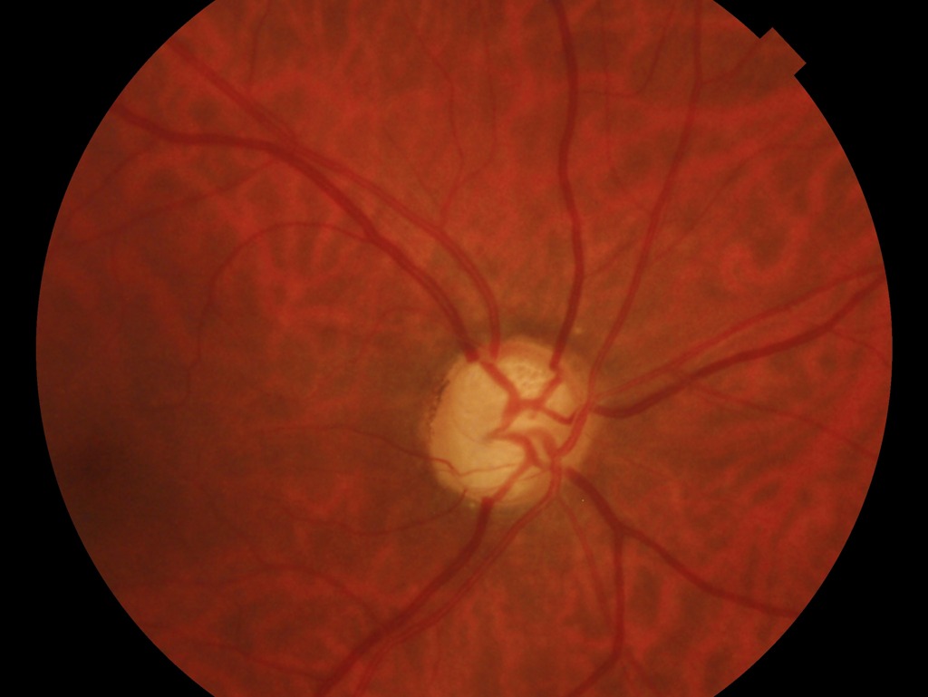
1. Find the disc
Stage A scans the whole photo
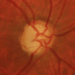
2. Crop around it
2.2 disc diameters, 256 × 256
3. Outline disc and cup
Stage B predicts two masks
4. Measure
vertical CDR = 0.74
Key facts
In GlaucomAI
Disc overlap (Dice) 0.98 on Chákṣu's test photos and 0.93 on PAPILA. Glaucoma AUC 0.85 on PAPILA and 0.72 on HYGD, cameras the model never saw.
Open the live demoReferences on this page
- 11.Ronneberger O, Fischer P, Brox T. U-Net: convolutional networks for biomedical image segmentation. MICCAI 2015, LNCS 9351:234–241. arXiv ↗
- 12.Kumar JRH, Seelamantula CS, Gagan JH, et al. Chákṣu: a glaucoma specific fundus image database. Sci Data. 2023;10:70. DOI ↗
- 13.Kovalyk O, Morales-Sánchez J, Verdú-Monedero R, et al. PAPILA: dataset with fundus images and clinical data of both eyes of the same patient for glaucoma assessment. Sci Data. 2022;9:291. DOI ↗
- 14.Abramovich O, Pizem H, Fhima J, et al. Hillel Yaffe Glaucoma Dataset (HYGD): a gold-standard annotated fundus dataset for glaucoma detection. PhysioNet, version 1.1.0. DOI ↗
- 15.Dice LR. Measures of the amount of ecologic association between species. Ecology. 1945;26(3):297–302. Google Scholar ↗